Durban Retinal AssociatesDurban Retinal Associates

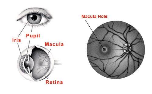
The retina is a thin layer of nerve tissue which lines the back of the eye.  The macula is the most sensitive portion of the retina and is responsible for the high definition central vision required for tasks such as reading, watching television, using a computer and driving.

A macula hole is a small hole in this area of the retina caused by degenerative changes in the vitreous, which is the jelly like substance which fills the back portion of the eye.  The symptoms of a macula hole are blurred central vision with or without some distortion.  You may battle to read with this eye and will often have difficulty in judging distances and may find activities like climbing steps or pouring a drink difficult.

SURGERY

VitrectomyA macula hole requires surgery to repair and this is performed by removing the vitreous (jelly) from the back of the eye (a vitrectomy) and filling the eye with gas, which will push the edges of the hole together and close the hole.  Surgical success (closure of the hole) is achieved in 90% of patients with one surgery.

After the surgery, the vision is initially poor because of the gas bubble in the eye which does not allow the light entering the eye to be focussed.  This gas bubble is gradually absorbed over a period of three to six weeks, and in this time, the vision will slowly improve.  Realistically one seldom gets a 100% visual recovery after this procedure, but the vast majority of people will notice an improvement in the quality of their vision and should have relief from symptoms such as distortion and difficulty in judging distances. The vision usually starts to improve from about 2 months after surgery and then takes approximately 12 months for complete healing to occur. The recovery (amount of visual improvement) depends on the size of the hole, the duration of the hole and each individual’s healing ability.

RECOVERY

After the surgery you will be required to posture in a specific position so that the gas bubble is centred over the hole and can adequately push the edges of the hole closed. In most cases one can sit or lie in any position (except lying flat on one’s back). This is important for the first 7 days after surgery and thereafter one can usually stop “posturing”.

It is also crucial that one does not travel to altitude and one does not fly in an aeroplane as long as there is a gas bubble in the eye.  Travel to altitude may result in a change in the pressure inside the eye as the gas bubble may expand and this can have catastrophic results.

The surgery is usually performed under general anaesthetic and requires a one night stay in the hospital.  It is sometimes possible to perform this procedure under local anaesthetic if there is a medical reason as to why you would not be suitable for a general anaesthetic or if you prefer a local anaesthetic.

One needs “bedrest” for approximately 1 week and then a slow return to one’s normal level of activity over the next three weeks avoiding strenuous physical exertion or vigorous exercise in this time. Walking (as an exercise) is possible from the 14 days following surgery. During the recovery phase there is no need to avoid using the operated eye and no possibility of overuse or “eye strain” of the fellow eye so reading, watching TV or using a computer are permissible. One only needs to keep the eye covered for the first 5 nights with a shield to prevent inadvertent rubbing. During the day the eye can left uncovered and you can wear your glasses as usual if required.

If the macular hole repair is combined with cataract surgery then one will need to change one’s glasses 3 to 6 months after the surgery depending on how quickly the vision recovers afterwards. If the surgery is done by itself then one may often not need a change of glasses. Your doctor will advise you on this during the follow up visits after surgery.

There is usually not much pain following the procedure.  Initially one may feel that the eye is a bit scratchy, but this usually settles very quickly.  The vast majority of procedures are performed using a sutureless technique which has dramatically helped speed up the recovery time.  The complications of this procedure include a retinal tear, a retinal detachment, cataract formation and an infection in the eye called endophthalmitis. Severe visual loss or blindness may occur as a complication in 1 in 10 000 cases These aspects will be discussed with you during the pre-operative consultation.